
A Retrospective Study Analysis of Data Trend of Erythema DyschromicumPerstans (EDP) Versus Lichen Planus Pigmentosus (LPP) Versus Post Inflammatory Hyperpigmentation (PIH) seen At DISHARC (DI Skin Health & Referral Centre) Nepal.
Introduction
In the recent years, there has been an increase in pigmentary disorders. Among those, hyperpigmentation is also quite common. When a patient visits dermatologist’s clinic with hyperpigmentation there are certain ways of asserting for the diagnosis. There are few hyperpigmentary disorders which are similar clinically and their proper diagnosis is required and further diagnostic skin punch biopsy is done as the treatment modalities, disease progression and several other factors vary. Nowadays, at the OPD clinic suspected cases of EDPvs. LPP vs. PIH are increasing. Hence a retrospective study of data was done to evaluate the various aspects of these pigmentary problems at DISHARC.
Discussion
Erythema dyschromicumperstans (EDP) is also known as ashy dermatosis. It is a benign, uncommon skin condition that presents with generalized ashy-grey colored macules on the body1. It affects both males and females, and is seen most commonly before the age of 40. Lesions still may be seen in young children and in the elderly.
Most patients are asymptomatic although some may have itching. On clinical presentation, the skin lesions are round, oval or sometimes irregularly shaped macules and patches that are grey, or bluish-brown in colour. They develop in a symmetrical pattern and can reach up to 0.5-3 cm in size. These lesions usually begin on the trunk and spread to the neck, arms and sometimes the face. Early lesions may have an elevated, non-scaly, reddish border and older lesions may have a lighter border. Mucous membranes are not affected.
A biopsy may be performed to rule out other diseases and skin conditions. There are a few therapeutic options for EDP but none that have been shown to be consistently effective. Clofazimine, an antibiotic with antibacterial and anti-inflammatory properties, and dapsone have been shown to be effective in some patients. Hydroquinone and topical steroids have varying results. Other therapeutic modalities include UV light therapy, chemical peels, griseofulvin, antihistamines, and isoniazid. EDP may persist for years although spontaneous remission may occur, especially in children.
Lichen planuspigmentosus (LPP) is a rare form of lichen planus. Areas that are exposed to sun such as the forehead, temples and neck are most commonly affected. However, the macules and patches may also develop on the trunk or in places where two areas of skin touch or rub together (i.e. the armpit, groin, etc).
Result:
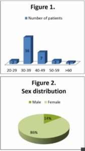
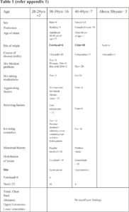
A single centre retrospective study over a period of 6 months was done at DISHARC. Data over a period of 6 months from year 2015 were collected and analysed.
After clinical diagnosis, the suspected cases were advised for skin punch biopsy and histopathological analysis for confirmation of the diagnosis.
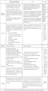
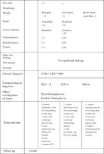
Morphologically lesion colour varied from ashy brown to blue gay and few were violet blue over the common site of origin which was usually over the chin followed by forehead and neck. 12 cases had well defined and 16 cases ill-defined border. There was absence of active red border, lichenoid papules, reticular pattern and none had pruritus and other skin findings. None of the cases were associated with nail, oral mucosa or genitalia findings.
Data were recorded in recording sheets and then evaluated (refer to appendix 1)
Performa EDP/LPP/PIH
Name Age/Sex
Address Profession
History:
Age of onset- Site of origin
Course of disease (months)
H/o any other medical problems –DM/ Htn./Hypothyroidism/ Hyperthyroidism/ Addison’s disease/ Others
H/o taking any medications
Aggravating factors
Relieving factors
H/o using cosmetics
Menstrual history
Clinical examination:
Distribution of lesion-
Localized (affecting one body area)
Generalised (affecting more than one)
Site: Forehead/ Cheeks (Rt./Lt./B/L) Chin/ Nose Symmetrical/ Asymmetrical
Neck Symmetrical/ Asymmetrical
Trunk
Chest Symmetrical/ Asymmetrical
Back (upper/ lower/ both) Symmetrical/ Asymmetrical
Abdomen Symmetrical/ Asymmetrical
Upper Extremities (upper arm/ forearm)
Lower extremities (upper leg/ lower leg)
Skin folds- Submammary folds, axillary, inguinal
Morphology of lesions:
Macules: Blue gray/ ashy brown/ violet blue
Border: well defined/ ill defined
Active red border: Present/ absent
Lichenoid papules: Present/ absent
Reticulate patter: Present/ absent
Pruritus: Present/ absent
Other skin findings-
Nail findings-
Oral mucosa-
Genitalia-
Clinical Diagnosis
Investigations:
Biopsy:
Hyperkeratosis/ hypergranulosis
Thinned epidermis
Basal cell vacuolization
Perivascular infiltrate
Incontinence of pigment & melanophages
Colloid bodies
Lichenoid infiltrate
Other changes
Histopathological diagnosis:
Other investigations (if done)
Treatment given
Follow up
References:
- org/page/Erythema Dyschromicumperstans
- 2011 Jan-Feb;9(1):63-4.A case of Cinderella: erythema dyschromicumperstans (ashy dermatosis or dermatosiscinecienta).Muñoz C1,Chang AL.
- JIMSA January-March 2014 Vol. 27 No.A Study of 10 Cases of Ashy Dermatosis and Lichen Planus Pigmentosum NeerjaPuri Consultant Dermatologist, Punjab Health Systems Corporation, Ludhiana, Punjab, India
- Fitzpatrik dermatology in general medicine Vol8Lowell A. Goldsmith, Stephen I. Katz, Barbara A. Gilchrest, Amy S. Paller, David J. Leffell, Klaus Wolff
- Lichen planus.DermNet NZ. March 2015; http://dermnetnz.org/scaly/lichen-planus.html.
- J ClinAesthetDermatol. 2010 Jul; 3(7): 20–31.PMCID:PMC2921758 Postinflammatory Hyperpigmentation A Review of the Epidemiology, Clinical Features, and Treatment Options in Skin of ColorErica C. Davis, MDa and Valerie D. Callender, MDa,b
*-*-*