
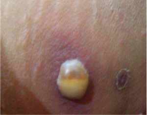
A 50 year old diabetic female was admitted with symptoms suggestive of angina pectoris. A dermatology reference was made in view of her cutaneous lesions. On enquiry, she had a history of slightly painful, multiple, pus filled lesions in her groin, lower abdomen and axilla, since 3 days. There was no history of fever. On examination, she had a few, non-tender, thin-roofed flaccid bullae filled with pus on an erythematous base, of size varying from 3 to 5 cm. These lesions showed a characteristic hypopyon formation. There were multiplesatellite follicular pustules as well. Healing lesions with collarette scaling were seen alongside (Figure 1). There was no regional lymphadenopathy. Fasting and post prandial blood glucose levels were 356 and 450mg/dl, respectively. Blood and urine ketone levels were normal.Her serum creatinine was 1.4 mg/dl. Hemogram with differential count was normal. Serum immunoglobulins and complement levels were normal. A pus culture revealed growth of Staphylococcus aureus phage type 55, sensitive to azithromycin, cloxacillin, methicillin, and cephalexin. A nasal swab showed no growth. She was started on Cephalexin 500mg twice a day and 2% mupirocin cream twice a day for a week which led to complete clearance of all the lesions.
Bullous impetigo is a bacterial pyoderma seen mainly in infants and young children. It is caused by group II phage types 55, 71 and 77 Staphylococcus aureus, which produce exfoliative toxins (ETs). ETs are serine proteases which cleave desmoglein directly 1and induce intraepidermal subgranular acantholysis, leading to blister formation. Hematogenous dissemination of ETs from a primary focus of infection can lead to the development of staphylococcal scalded skin syndrome (SSSS). The rarity of these disorders in adults is primarily due to a superior capacity to metabolize and excrete the toxin, as well as more efficient immune capabilities.2Diabetes mellitus is associated with an enhanced risk of infections, sometimes with an increased severity. Some of these may be potentially serious and associated with a greater risk of complications.3 Staphylococcus aureus infections may occur with greater frequency among patients with diabetes mellitus.4 Bacterial infections can worsen glycemic control and lead to diabetic ketoacidosis.5
This case illustrates how impaired host resistance can lead to the development of uncommon types of bacterial infections in adults. She had a high blood sugar level with poor renal function which probably led to the characteristic manifestation seen normally in children. Such patientsare at an increased risk of developing SSSS.
References:
1) Yasushi Hanakawa, John R. Stanley. Mechanisms of Blister Formation by Staphylococcal Toxins. J Biochem 2004;136(6):747-750.
2) Peter M. Elias, Peter Fritsch, Ervin H. Epstein, Jr. Staphylococcal Scalded Skin Syndrome: Clinical Features, Pathogenesis, and Recent Microbiological and Biochemical Developments. Arch Dermatol 1977;113(2):207-219.
3) Nirmal Joshi, Gregory M. Caputo, Michael R. Weitekamp, A.W. Karchmer. Infections in Patients with Diabetes Mellitus. N Engl J Med 1999;341:1906-1912.
4) Breen JD, Karchmer AW.
Staphylococcus aureus infections in diabetic patients. Infect Dis Clin North Am 1995;9(1):11-24.
5) Elie Azoulay, Sylvie Chevret, Judith Didier, Segolene Neuville, Michel Barboteu, Caroline Bornstain, et al. Infection as a Trigger of Diabetic Ketoacidosis in Intensive Care—Unit Patients.Clin Infect Dis 2001;32(1):30-35.