GST Implications for a Dermatologist- A Session on GST at CDCON 2017

CDCON 2017

Among the so many useful derma conferences being organised in India, it is a big challenge for the organisers to sustain with newer sessions and practical useful content, at CDCON we are no different but a dedicated team of 40+ Dermatologists striving to make it a NOT TO MISS CONFERENCE and in three years time we are encouraged to make it to a multi-state level and raise the bar. CDCON 2017, a conference for Dermatologists, was held during September 09-10, 2017 at Ahmedabad. We aptly defined it as “a conference designed to uplift your practice to the next level” because there were insightful sessions on significant issues pertaining to the domain of Dermatology. Spread over two days, the Conference addressed some of the key aspects of Dermatology practice. The sessions were conducted by some of the leading Dermatologists in the field.

While double chin correction with Deoxycholic Acid fascinated the audience with actionable ideas and interesting case studies, practical sessions on marketing and
up lifting practice also WOWed the audience. A whole dedicated day allotted to Dermatologists’ staff Soft Skills Development and Motivational training became talk of the town among the delegates, as the efficiency and effectiveness of support staff have a bearing on the overall functioning of a clinic and as such training is of vital significance for every Dermatologist’s practice.

All scientific sessions followed by very interactive and knowledge sharing panel discussions at CDCON, as we strongly believe the best knowledge would be shared out of interactive Q & A and panel discussion. Presentations on ‘Art of Communication & Counselling’, ‘Tips to Buy Laser Machines’, ‘Smart Marketing’ and ‘How to Balance Aesthetic and Clinical Practice’ were quite enlightening and interesting.

Among all the interactive-practical important sessions, I found the session on
Goods and Services Tax by Mr. Nilesh Suchak on Day 2 particularly informative and useful and I thought all the dermatologists should be benefited by that GST session what better way could be to share the key points of the session by sharing it to medium of DERMATALK magazine. Here are the key points:

The session on GST addressed many of the queries that Dermatologists had. As our schedule as a Dermatologists are quite hectic and do not permit sufficient time to study the implications of GST for us, the session on GST helped clarify some of the assumptions and queries. The presentation on GST dwelt upon the structure of GST and the way it is calculated. It clarified as to when the GST Registration becomes a mandatory requirement.

While ‘healthcare services’ are included in the services ‘exempt’ from GST, it was interesting to learn about the way GST defines ‘healthcare services’. It is important to study this definition as it has implications for Dermatologists. Healthcare services are defined as follows:

“health care services” means any service by way of diagnosis or treatment or care for illness, injury, deformity, abnormality or pregnancy in any recognized system of medicines in India and includes services by way of transportation of the patient to and from a clinical establishment, but does not include hair transplant or cosmetic or plastic surgery, except when undertaken to restore or to reconstruct anatomy or functions of body affected due to congenital defects,
developmental abnormalities, injury or trauma.

As it is clear, the treatments such as hair transplant or cosmetic or plastic surgery are not included except in the special circumstances/conditions as mentioned in the clause. In addition, terms such as ‘clinical establishment’ have also been defined for Dermatologists to study them in detail. The presentation went on to enlist the kind of returns are to be filed in the event of an excess to the aggregate turnover. GST has clearly articulated the classification of healthcare services which affects the community of Dermatologists and should be studied in great
detail.

The presentation listed the following scenarios for the same:

  • When aggregate practice exceeds Rs. 20 lakhs, a dermatologist should take GST Number.
  • It is a dual-tax method-CGST + SGST – 9% each, total 18%

1. It is clear from above discussion that all dermatologists who are doing only consultation & clinical practice and their annual income is below 20 Lakhs, need not apply for GST number, but if it crosses 20 Lakhs, one has to take GST number. Once GST number is applied they are supposed to file a return, though their income is exempted under health care service – nil return is a must in this case.
2. All those dermatologists who are doing MIX PRACTICE – Dermatological & Cosmetological, they are supposed to take GST Number, irrespective of their annual income.

3. Once GST Number is applied and taken then they are supposed to file 37 RETURNS annually, 3 returns monthly and 1 annually.
4. There is a special proforma to generate GST bill & it should be printed and changed accordingly.
5. Procedures which are because of medical diseases are exempted from GST. As they are included under “Health Care Services”, like wart removal.
As a Dermatologist, it was an enriching experience to not only attend the conference but also acquire an in-depth understanding and knowledge of GST and its implications on my practice. While it is not possible to fully internalize all legal implications of GST at present, it is at least clear to me, thanks to the session on GST, how GST will affect my practice and how I need to make the transition
to the new taxation system namely GST. In a one-hour presentation, it was truly possible for me to experience how I can take my Dermatology “practice to the next level” with respect to GST, as the Conference tag line goes…

*-*-*

Nanotechnology in Dermatology

What is Nanotechnology?

Nanotechnology can be described as a collection of methods and techniques for processing materials at an atomic and molecular scale to create products with special physicochemical properties such as rigidity, hydrophobicity, size, shape, charge, conductance, chemical reactivity, magnetism in relation to conventional products. It deals with materials less than 100 nm. There are two major approaches to get the nano particles -one is the bottom up and the other is top down approach. Top-down fabrication reduces large pieces of materials all the way down to the nano scale while bottom-up approach to nano manufacturing creates products by building them up from atomic- and molecular-scale components.

History of Nanotechnology:

The idea and concept behind nanotechnology started with a talk entitled “There’s Plenty of Room at the Bottom” by physicist Richard Feynman at an American Physical Society meeting at the California Institute of Technology (CalTech) on December 29, 1959, long before the term nanotechnology was used. Feynman described a process in which scientists would be able to manipulate and control individual atoms and molecules. Over a decade later, Professor Norio Taniguchi coined the term nanotechnology, in his explorations of ultraprecision machining. In 1981, with development of the scanning tunneling microscope that could see individual atoms by IBM and the modern nanotechnology era began.

Type of Nanoparticles:

Nanoparticles fall into 3 categories:

1) Naturally occurring: particles constantly present in nature such as ashes produced from volcanoes, dust, nanoparticles produced from water bodies and plants, viruses etc.

2) Incidental: It includes combustion products, industrial processes, vehicle emission, from construction process, welding fumes etc.

3) Engineered: They are formed for human welfare and divided into organic and inorganic.

Organic particles namely lipososmes, dendrimers, fullerenes, PAMAMS, ISCOMS, polymerised particles, solid lipid nanoparticles, nanostructured lipid carriers.

Liposomes:

Liposomes are biocompatible, versatile and have good entrapment efficiency. It finds application as long circulatory delivery of gene, drug, protein and peptide.

Fullerenes:

They are 1-nm carbon spheres of 60 carbon atoms and capable of carrying genes, proteins and other biomolecules for delivery purposes.

Dendrimers:

They are hyper branched, tree-like structures. Unimolecuar structure with size of 20nm & used to transport medication.

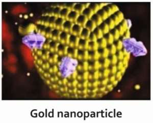
Inorganic nanoparticles are made of metal (gold or silver), metal oxide (iron), and ceramic material (silica), encapsulating medication in their interior, transferring and liberation various substances, quantum dots and carbon tubes.

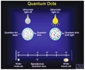
Quantum Dots are 2-10nm nanocrystalline semiconductor material made from cadmium selenide with an exclusive spectroscopy and with optical properties good candidates for diagnostic applications.

Carbon nano tubes: These are unique for their size, shape, and have unique physical properties. Nano tubes have some special advantages over other drug delivery and diagnostic systems due to their unique physical properties.

Applications of Nanotechnology in Dermatology:

Broadly classified into 3 categories:

  1. Cosmeceuticals: sunscreens, volatile compounds like insect repellents, perfumes, emollients, anti-wrinkle creams.
  2. Diagnostic devices: Carbon nanotubes, Quantum dots, Nanopunches
  3. Therapeutic devices:As  drug delivery carriers, or nanosized drug itself.

1) Cosmeceuticals 

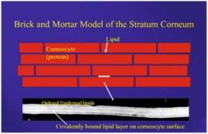
Nanoparticles are used to encapsulate a vast range of substances beneficial to skin. In this way nanovesicles are developed for both pharmaceutical and cosmetic use. They are applied over the skin and these agents enter either through the upper layer of skin known as stratum corneum or through the hair follicles.

Sunscreens are one of the most commonly used over-the-counter drugs. Nanoparticles  of titanium dioxide and zinc oxide are used in sunscreens which increases the efficacy, decreasing the white effect remain after using the conventional ones and decreases their stickiness  and  covering the skin more evenly. While use of nanoparticles in moisturisers increases their permeability into skin.

Use of botulin toxin and hyaluronic acid used as anti-ageing agents. Hyaluronic acid in its bulk form is 50,000 nm or larger in size and is unable to penetrate skin when applied topically. While nanosized particles of hyaluronic acid can penetrate the skin and are the basis of a topical delivery system in development. Botulinum toxin has been stabilized and encapsulated in a form that allows penetration of the skin and apparent effacement of rhytides in early clinical trials.

Chitin nanofibrils are natural polysaccharide obtained from crustacean shells after carbonate and protein removal. These nanofibrils activate keratinocyte and fibroblast proliferation, cells present in skin, and regulate collagen synthesis.

Other uses like slow release kinetics are important for perfumes, which can yield all-day fragrance. They are also useful for insect repellents, such as N, N-Diethyl-meta-toluamide (DEET), to prolong efficacy. Nano-emulsions provide a new colloidal drug delivery system that efficiently replaces lost lipid in epidermis.

2) Therapeutic:

A) As a drug delivery system:

Used as carrier for different drugs with benefits of site specific drug delivery, lesser side effects, more duration of action of the drug due to sustained release, protecting the therapeutic agent from chemical, physical and biological damage and increased cellular uptake of drug.

Thus variety of nanoparticles such as dendrimers, lipososmes, polymeric nanoparticles, solid nanoparticles, nano structured lipid particles etc are being used as drug delivery system. They encapsulate the drug and deliver it to the desired site.

Drugs which are commonly used nowadays with nanotechnology are analgesics with their sustained release properties, antibiotics covered with polymeric nano particles, cortico-steroids decreasing inflammatory process inside the body, drugs for hair fall, acne treatment, anti-fungals, silver used as an anti bacterial agent in wound dressing and many agents are being used in many other diseases.

Antisepsis is another big area for nanoparticle operation. Chlorhexidine gluconate carried by nanoparticles (Nanochlorex®) is having an immediate antibacterial effect. The most commercialised antibacterial nanomaterial to date is nanosilver, used not just in wound and burn dressings but also as water disinfectant and room spray.

B) In treatment of malignancies:

Due to the small size of nano particles can be of great use in oncology, both in diagnosing and treating malignancies.  Nano shells coated with gold were used to kill cancer tumors in mice by Prof. Jennifer at Rice University. These nano shells are targeted to bond to cancerous cells by conjugating antibodies or peptides to the nano shell surface. Area of the tumor is irradiated with an infrared laser, which heats the gold sufficiently and kills the cancer cells.

nanotechnology

Nanoparticles are used in cancer photodynamic therapy, wherein the particle is inserted within the tumor in the body and is illuminated with photo light from the outside. The particle absorbs light and if it is of metal, it will get heated due to energy from the light. High energy oxygen molecules are produced due to light which chemically react with and destroy tumors cell, without reacting with other body cells. Photodynamic therapy has gained importance as a non-invasive technique for dealing with tumors.

C) Gene Silencers:

Gene silencing is currently one of the most promising new approaches for disease therapy. This is useful in genetically inherited diseases or diseases occurring due to mutation in genes. Small interfering RNA (siRNA) showed significant potential in new molecular approaches to down-regulate specific gene expression in mammalian cells. However, RNAs cannot easily penetrate cell membranes; therefore RNA delivery becomes one of the major challenges for gene silencing technology. Small interfering ribonucleic acids encapsulated with polymeric nanoparticles can precisely inactivate the gene expression and hence useful in genetic disorders.

3) Nano-diagnosis:

One of the important uses of nanotechnology is directed towards development of improved diagnostic techniques to screen complex diseases. Screening indicates identification of the cause of illnesses, monitoring the improvement or progression of the state of diseases such as cancer, cardiovascular or neurodegenerative diseases.

Nanotechnology enables the manipulation of materials at nanoscale and has shown potential to enhance sensitivity, selectivity and lower the cost of diagnosis. Several types of nanomaterials such as metals, metal-oxides and quantum dots have shown ample advantages over traditional diagnosis.

Quantum dots, with quantum confinement properties, can be used in conjunction with MRI, to produce exceptional images of tumor sites. Fluorescent quantum dots produce a higher contrast image and at a lower cost than organic dyes used as contrast media. It works on the principle of absorbing light of desired wavelength and having envelope with fluorescence signal. When tagged with antibodies they emit fluorescence that helps in diagnosing  tumours

In malignant melanoma, topical application of quantum dots will allow sentinel lymph node evaluation without disturbing the skin and used as a non-invasive method to evaluate the spread of malignancy.

Gold nanoparticles, in combination with fluorescent protein, are being used in diagnosis of cancer by emitting fluorescence when binding occur with the target atypical cell.

nanotechnology

Carbon nanotubes 0.5–3 nm in diameter, are specialised structures having good conductivity with special property of alteration in their conductivity on binding to macromolecules like nucleic acids and antibodies, their conductivity changes. They are used in diagnosis of skin infection and malignancies, for detection of DNA mutation.

Nanopunches are small simple biopsy tool that is made from silicon, chromium, nickel & copper. Having unique shape of ‘órigami claw’ like, its movement and excretion controlled by magnetic field and useful to obtain biopsy from difficult sites like nail matrix, fascia, liver etc.

Iron oxide nanoparticles can used to improve MRI images. The magnetic property of iron oxide enhances the images from the Magnetic Resonance Imagining scan.

Adverse Effects:

1) Formation of foreign body granuloma

2) Scleromyxedema i.e hardening of organs

3) During pregnancy can be hazardous for the foetus

4) Free radical injury to the cells by particles TiO2

5) Act as hapten or cross reactant and can start allergic reactions

Nanoparticles are present in our atmosphere since the origin of earth, for example as the result of combustion processes (forest fires), in volcanic ash, or as dust in the air due to weathering and erosion. In the modern era, they are unintentionally produced due to human activities like household heating, industry, slash-and-burn clearance, transport. All these things adversely affect the atmosphere and the creatures but no studies are available that can explain the mechanisms of uptake, distribution, metabolization and excretion of nanoparticles and exact damage they are causing to environment.

Advantages of Using Nanotechnology:

1) Increases efficacy of drugs

2) Provides a site specific drug delivery therefore reducing uneventful side effects

3) In cosmetology, having many indications and used in anti-ageing process

4) It improves the diagnosis and treatment of many diseases

5) As a gene therapy can prevent the inherited diseases

6) Nano-built devices can be implanted in body to continuously adjust body’s chemical balance

There is a never ending list of benefits delivered by nanotechnology in different fields apart from health and medicine. It gives a new way of providing healthcare to humans.

References:

Nasir A : Nanotechnology and dermatology: part 1 –potential of nanotechnlogy. Clin Dermatol. 2010;28(4) : 458 -66

Ali R , Hussain I –Nanoderamtology : new horizon . J Pak Assoc Dermatol.2013;23(2):117-9

Newman MD, Scotland M , Elis JI . The safety of nanosized particles in titanium dioxide and zinc oxide based sunscreens . J Am Acad Dermatol. 2009;61 (4):685-92.

Nanodermatology society. Available from: http ://www.nanodermsociety.org/index.php.

Vitiligo Minigrafting over Difficult Areas

Abstract

Vitiligo is a result of disturbed epidermal melanization with an unresolved etiology and incompletely understood pathogenesis.

Various treatment options have resulted in various degrees of success. Various surgical modalities and transplantation techniques have evolved during the last few decades. Of them, miniature punch grafting (PG) has established its place as the easiest, fastest, and least expensive method. Various aspects of this particular procedure, especially over the difficult areas have been discussed here.

A detailed discussion on the topic along with a review of relevant literature has been provided in this article.

Key Words: Mini grafting, mini punch grafting, punch grafting, vitiligo, vitiligo surgery

Introduction

Many patients respond to standard medical treatment options. However, several patients remain recalcitrant or respond only partially. Any attempt to repigment these resistant patches with conventional medicinal modalities is often unsuccessful and sometimes frustrating, indicating the absence of a melanocytes reservoir, to induce repigmentation. Under these circumstances, melanocyte repopulation of the achromic areas is not possible, unless a new source of pigment cells is placed by surgical methods within the depigmented lesion/s.

Different corrective surgical methods have evolved during the last four decades. Some of these are: Thin Thiersch’s graft, [1] epidermal grafting by suction blister, [2] punch grafting, [3] mini punch grafting, [4],[5] cultured melanocyte grafting, [6],[7] grafting of cultured epidermis, [8]autografting, and PUVA, [9,10] transplantation of autologous cultured melanocytes, [11] single hair transplant, [12] ultrathin epidermal sheets and basal cell layer suspension [13] and mini grafting and NB-UVB. [14]

Among all these, mini punch grafting (PG) is the easiest, fastest, least aggressive, and a technique with minimal expenses.[14]

Test Grafting

Before embarking upon any surgical intervention in vitiligo, a proper assessment of the stability status is of paramount importance. In recent times, this concept has been discussed in detail. [15]


Clinically, stability can be judged by three simple indicators:

  • History
  • Lack of progression of old lesions and absence of development of any new lesion within a specified period (6 months to 2 years)
  • Koebner phenomenon (Kp)
  • Absence of a recent Kp either from history (Kp-h) or experimentally induced (Kp-e)
  • Test grafting

On the backdrop of persistent incongruity about the minimal period of stability, an attempt was made for the first time by Falabella, in 1995, to fathom the stability before surgery, by introducing a mini grafting test. [16]

The objective of this test was to:

  • Establish the stability of the depigmenting process
  • Determine a means by which patients could be selected
  • Identify patients who may respond to pigment cell transplantation
  • Anticipate the response to surgical repair

In the original suggested procedure, a few grafts (1.0-1.2mm) were placed in the centre of the depigmented lesion to be scrutinized. Dressing was done by Micorpore® adhesive tape and kept for a couple of weeks. After removal of the tape, the area was exposed to sunlight for 15 minutes daily, for a period of 3 months. No treatment was permitted during this test period.

All test sites were visualized under Wood’s light. The test was considered positive if unequivocal repigmentation took place beyond 1mm from the border of the implanted grafts. On the other hand, if less than 1mm or no repigmentation was observed the test was considered to be negative.

In some of the biggest series, this evaluation has been termed as ‘test grafting’ (TG) and is found to be a more reliable exercise than the unjustified dependence on the period of stability alone. [14],[17],[18]

Over the years, this ‘test’ has been vindicated and acknowledged as a powerful tool for detecting stable vitiligo, which anticipates the repigmentation success in vitiligo when surgery becomes a therapeutic option.

After proper assessment of the stability status and routine physical examination and investigations, an informed consent is taken from the patient, and the donor and recipient areas are surgically prepared.

  1. The instruments required are 1.5 or 1.2mm punches, small jeweler’s or graft holding forceps, and a small curved tip scissors.
  2. The recipient area is prepared first. Two percent lignocaine with or without adrenaline is infiltrated as a local anesthetic.
  3. To minimize the chance of developing any perigraft halo, the initial recipient chambers are made on or very close to the border of the lesion. The punched out chambers are spaced according to the result of test grafting or at a gap of 5-10mm from each other.
  4. The donor area is either the upper lateral portion of the thigh or the gluteal area. Punch impressions are made very close to each other so that a maximum number of grafts can be taken from a small area.
  5. Same sized punches are used for both the donor and recipient areas.
  6. The grafts are placed directly from the donor (buttock/upper thigh) to the recipient areas. This speeds up the procedure and lessens the chance of infection. Care is taken to ensure that the graft edges are not folded and the tissue is not crushed or placed upside down. The needle of the syringe or the tip of the scissors is used for the proper placement of grafts in the recipient chambers.
  7. Hemostasis is achieved by pressing a saline-soaked gauze piece over the area.
  8. For the recipient area, the three layers of dressing from inside to out are: Paraffin-embedded, non-adherent sterile gauze (Jelonet®), sterile Surgipad® , and bio-occlusive Micropore®.
  9. For the donor area, only Surgipad®and Micropore® are used.
  10. The recipient area may be immobilized if necessary. Proper instructions for special areas like the lips are necessary. To secure the recipient area, these patients are advised to be on a liquid diet for the first 24 hours, preferably with a straw. Patients are allowed a normal diet after this period.
  11. Sometimes dressings are opened after 24 hours to look for any dislodgement of grafts. If any are found, they are replaced.
  12. Finally, after 4 to 7 days, the dressings are removed.[17],[19],[20],[21] [Fig-]

Follow-up and Course of Events

Post-surgery, the patients are exposed to PUVA [9],[10] / PUVASOL (Psoralen plus UVA from Sunlight) [17],[21] or NB- UVB [14] or even kept as such in some studies. [17] The patients are followed up fortnightly for the initial two months and then monthly, until complete repigmentation is achieved.

In the donor site, after healing with secondary intention, minimal superficial scarring is expected and acceptable.

Scabs may fall off from the recipient site within 7-14 days. However, in many instances, there may not be any scab formation. Perigraft repigmentation is expected to start from around 3-4 weeks. [14],[17],[21]


The entire depigmented and grafted area is expected to be completely repigmented within 3-6 months, based on the area of grafting and the body part involved.

In this article, miniature punch grafting was displayed over upper lip (Fig 2a-2d), eyelids (Fig 3a-3b, 4a-4b) (both upper and lower), male genitalia (Fig 5a-5f), external ear (6a-6b, 7a-7b), nipple (8a-8d).

Complications

Recipient site

  1. Cobble stoning
  2. Polka dot
  3. Variegated appearance and color mismatch
  4. Static graft (no pigment spread)
  5. Depigmentation of graft
  6. Perigraft halo
  7. Graft dislodgement/rejection
  8. Hypertrophic scar and Keloid formation

Donor Site

  1. Keloid
  2. Hypertrophic scar
  3. Superficial scar
  4. Depigmentation/spread of disease
  5. Contact dermatitis to adhesive tapes

By proper selection of cases most of these complications are entirely avoidable. [5],[19],[20],[21]

Cobble stoning is regarded as the commonest of them all. [14],[17],[18],[21] 
It was observed that with time it got corrected in most of the cases. [17] In resistant cases, corrective electrofulguration may be needed. [22]

In this regard it is only apt to conclude that grafting should not be performed with punches more than 1.5mm in diameter. On face and lips, it should be even smaller (1.2mm or 1mm). [14],[23] 


Herpes labialis-induced lip leucoderma (HILL) is another unpredictable entity bearing the risk of rejection of grafts. [24],[25],[26],[27]

Advantages

  1. Easiest, fastest, and least expensive method
  2. High rate of success with very few preventable/manageable side effects
  3. Can be performed anywhere, on any site (except angle of the mouth)

Discussion

Surgical correction of vitiligo and other cutaneous achromia have come a long way in the last five decades or so.

However, among all other methods, autologous miniature punch grafting has established its place as the easiest, fastest, safest, and least aggressive means of vitiligo surgery.

When the graft is taken off, the piece of tissue is completely detached from the donor site and then placed on the vascular bed in the recipient holes. From this vascular bed, it derives its blood supply. Initially, the graft adheres to its new bed with the help of fibrin. There is diffusion of nutrients through this fibrinous layer, which keeps the graft alive initially. Within 2-3 days, capillary linkage occurs, with vascularization of the graft. The thinner the graft the denser the capillary network in the superficial dermis, and thus earlier is the process of vascularization. [28]

Acknowledgment

Some figures and tables used in this article have also been used in the following chapter and articles by me and my co-authors.

I would like to properly acknowledge the original source:

  1. Lahiri K, Malakar S, Sarma N, et al. Repigmentation of vitiligo with punch grafting and narrow-band UV-B (311 nm) a prospective study. Int J Dermatol.2005;45(6):649-55.
  2. Malakar S, Lahiri K. Spontaneous repigmentation in vitiligo: why it is important? Int J Dermatol. 2006; 45:477-8.
  3. Malakar S, Lahiri K. Mini grafting for vitiligo. In: Gupta S, Olsson MJ, Kanwar A J, Ortonne JP (Eds).Surgical Management of Vitiligo, 1st edition. Oxford: Blackwell Publishing; 2007;87-95.
  4. Lahiri K.  and evaluation of autologous mini punch grafting in vitiligo. Indian J Dermatol. 2009;54: 159-167
  5. Lahiri K.Autologous Mini Punch Grafting in Vitiligo. In: Lahiri K, Chatterjee, Sarkar R(Eds). Pigmentary Disorders A Comprehensive Compendium , 1st Jaypee brothers; 2013;241-250

References

1. Behl PN. Homologous thin Thiersch’s grafts in treatment of Vitiligo. Curr Med Pract 1964;8:218-21.
2. Falabella R. Epidermal grafting: An original technique and its application in achromic and granulating areas. Arch Dermatol 1971;104:592-600.
3. Orentriech N, Selmanwitz VJ. Autograft repigmentation of leucoderma. Arch Dermatol 1972;105:784-6.
4. Falabella R. Repigmentation of leucoderma by minigrafts of normally pigmented, autologous skin. J DermatolSurgOncol 1978;4:916-9.
5. Falabella R. Repigmentation of segmental vitiligo by autologous mimigrafting. J Am AcadDermatol 1983;9:514-521.
6. Lerner AB, Halaban R, Klaus SN, Moellmann GE. Transplantation of human melanocytes. J Invest Dermatol 1987;89:219-24.
7. Lerner AB, Repopulation of pigmented cells in patients with Vitiligo. Arch Dermatol 1988;124:1701-2.
8. Falabella R, Escobar C, Borrero I. Treatment of refractory and stable vitiligo by transplantation of in vitro cultured epidermal autografts bearing melanocytes. J Am AcadDermatol 1992;26:230-6.
9. Skouge JW, Morison WL, Diwan RV, Rotter S. Autografting and PUVA: A combination therapy for vitiligo. DermatolSurgOncol 1992;18:357-60.
10. Hann SK, Im S, Bong HW, Park YK. Treatment of stable vitiligo with autologous epidermal grafting and PUVA. J Am AcadDermatol 1995;32:943-8.
11. Olsson MJ, Juhlin L. Repigmentation of Vitiligo by transplantation of cultured autologous melanocytes. ActaDermVenereol (Stockh) 1993;73: 49-51.
12. Malakar S, Dhar S. Malakar RS. Repigmentation of vitiligo patches by transplantation of hair follicles. Int J Dermatol 1999;38:237-8.
13. Olsson MJ, Juhlin L. Long-term follow-up of leucoderma patients treated with transplants of autologous cultured melanocytes, ultrathin epidermal sheets and basal cell layer suspension. Br J Dermatol 2002;147:893-904.
14. Lahiri K, Malakar S, Sarma N, Banerjee U. Repigmentation of vitiligo with punch grafting and narrow-band UV-B (311 nm) a prospective study. Int J Dermatol 2005;45:649-55.

15. Malakar S, Lahiri K. Minigrafting for vitiligo. In: Gupta S, Olsson MJ, Kanwar AJ, Ortonne JP, editors. Surgical management of vitiligo. 1 st ed. Oxford: Blackwell Publishing; p. 87-95.

16. Falabella R, Arrunategui A, Barona MI, Alzate A. The minigrafting test for vitiligo: Detection of stable lesions for melanocyte transplantation. J Am AcadDermatol 1995;32:228-32.

17. Malakar S, Dhar S. Treatment of stable and recalcitrant vitiligo by autologous mimiaturepunch grafting: A prospective study of 1,000 patients. Dermatology 1999;198:133-9.

18. Malakar S, Lahiri K. Punch grafting for lip leucoderma. Dermatology 2004;208:125-8


  1. Falabella R. Repigmentation of segmental vitiligo by autologous mimigrafting. J Am AcadDermatol 1983;9:514-21.
20. Malakar S. Punch Grafting. In: An Approach to Dermatosurgery. 1 st ed. Calcutta: A Paul, 1996. p. 44-6.
21. Lahiri K, Sengupta SR. Treatment of stable and recalcitrant depigmented skin conditions by autologous punch grafting. Indian J DermatolVenereolLeprol 1997;63:11-4.
  1. Malakar S, Lahiri K. Electrosurgery in cobblestoning. Indian J Dermatol 2000;45:46-7.
  2. Falabella R. Surgical treatment of Vitiligo: Why, when and how. J EurAcadDermatolVenereol 2003;17:518-20.
24. Malakar S. Dhar S. Rejection of punch grafts in three cases of herpes labialis induced lip leucoderma, caution and precaution. Dermatology 1997;195:414.
25. Malakar S, Dhar S. Acyclovir can abort rejection of punch grafts in herpes-simplex induced lip leucoderma. Dermatology 1999;199:75.
26. Malakar S. Lahri K, Successful repigmentation of six cases of Herpes labialis induced lip leucoderma by micropigmentation. Dermatology 2001;203:194.
27. Lahiri K, Malakar S. Herpes simplex induced lip leucoderma: Revisited. Dermatology 2004;208:182.
  1. Burge S, Rayment R. Free skin grafts. In Simple skin Surgery. 1st Mumbai: Blackwell Scientific Publications; 1986. p. 71-84.


Complications Of Filler And Neuromodulator Injections

Experience teaches aesthetic practitioners to become good, but avoiding and managing complications make them great.  Although complications related to dermal filler and toxin injections remain rare, understanding what can go wrong and how it can go wrong are essential when we’re using these products. A well-trained practitioner is able to reduce the sequelae from an adverse event by acting promptly using algorithms and a methodical approach to treatments.

Fillers           

Complications are best prevented with careful planning. Thorough knowledge of both anatomy and the specific characteristics of each filler are critical. The appropriate depth of placement is dependent on the product selected and strongly influences the end result. In order of depth, medium hyaluronic acid products should be injected into the deeper dermis, calcium hydroxyapatite at the dermal-subcutaneous border, and poly-L-lactic acid in fat below the dermis. An injection placed too superficially can produce nodules and an uneven surface; therefore, it is usually preferable to err on the deeper side. 

Before treatment, the physician should clarify the patient’s goals and expectations for both the procedure itself and the subsequent results, and the patient should be given the opportunity to identify desired treatment areas with a mirror. Standardized pre- and post procedure photographs should be taken for documentation. It’s essential to obtain informed consent; this should cover the discussion of potential side effects and all details of pre and postoperative care. The patient should be screened for any medical issues that could affect the application of filler, such as pharmacologic or pathologic clotting disorders, previous vagal episodes, and any history of seizures.

Patients are warned to avoid NSAISs, aspirin, vitamin C, and omega 3 supplements. I usually suggest arnica tablets to reduce bruising risks. It is recommended that patients sleep with the treated area elevated for 1 to 2 nights after injections. Ice water bags applied to treated areas for 5 to 10 minutes per hour will reduce swelling/edema and trauma. I always apply a topical cooling device that reduces pain and bruising after applying topical anesthetic agent before the injections. Using a cannula will reduce bruising. When a bruise appears, it is useful to offer topical applications or sometimes vascular laser treatment to speed resolution. It is well documented that there is a direct correlation between the speed of injections and the number of complications, so it is essential to decrease the speed of injections; I usually spend 5 to 10 minutes per ml of filler, depending on the area injected. Using the smallest gauge needle also slows down the administration of filler. In the event of “lumps and bumps” massage is helpful with many types of fillers, but not all, if they cannot be massaged away after 7 to 10 days, it is possible to use hyaluronidase. It is also possible to extrude many types of filler by using a puncture with a 26-gauge needle or an 11 blade. These same techniques may also be utilized for areas that have been over corrected.

Granulomas are more commonly seen with non hyaluronic acid based products, these may form microspheres that can be extremely difficult to treat. Treatment with hyaluronidase, collagenase, and steroids has demonstrated to help these granulomas. In some instances of granulomas, surgical removal is the definitive treatment. When granulomas undergo delayed inflammation, treatment with oral antibiotics and cyclooxygenase-2 inhibitors may help to minimize the swelling. When injecting the neck and chest, the use of the microdroplet distribution of hyaluronic acid–based products may help to decrease the risk of granulomas. Hyaluronidase is used at 20 to 60 units per 1-mL filler. Triamcinolone is injected to females (2.5 mg/mL) and males (5 mg/ mL) with 0.1 mL per granuloma. The use of calcium hydroxylapatite on the dorsum of the hands could often lead to swelling, and to minimize this I usually place 1 to 1 ½ cc split between both hands and the additional filler is placed at a follow up visit 1 month later if needed.

Infectious complications are another risk of filler injections and can include herpes simplex infection with or without secondary bacterial infection, acute bacterial infections, or biofilm-related events. Practitioners should be using a prep that is effective in treating both gram-positive and gram-negative bacteria. An abscess at a single site suggests contamination was introduced during the injection, whereas the appearance of multiple abscesses indicates the patient was injected with filler material that may likely have been contaminated prior to injection. Sometimes the anesthetic mixed with filler becomes the source of contamination. It’s an option to administer fillers that come pre-mixed with an anesthetic to reduce the risk of contamination. Multi-dose vials of anesthetic should be dated once opened and unused portions should be discarded to reduce the risk of contamination. Also it is not a best practice to store partially used syringes of product for reinjection later. Recurrent abscesses as well as delayed foreign body granulomas are probably related to biofilm, which represents a structured community of micro-organisms encapsulated and protected within a self-developed polymeric matrix, which adheres irreversibly to host tissue or an implanted device. Needle drainage should be performed on any fluctuant lesion and the aspirated material should be sent for culture or PCR analysis. Empiric antibiotic treatment should be initiated with a quinolone plus a macrolide, but the regimen can be altered as dictated by the microbiology results. If the lesion does not respond to antibiotic treatment and hyaluronic acid filler was used, hyaluronidase injection can be performed. As a last resort, consideration can be given to use of pulsed intralesional steroids, once s week for two or three weeks. Biofilm-related complications of filler injections are probably not completely preventable but there are strategies that can be followed to minimize the risk like avoiding injections in the perioral area in patients with an active infection as well as any use of an intraoral injection route. Avoid applying makeup prior to the procedure and for eight hours afterward, and the practitioner should adhere carefully to aseptic injection technique to reduce the bacterial load, I prefer to use chlorhexidine to prepare the skin because unlike alcohol, it has a residual antibacterial effect. Using a smaller gauge needle will minimize surface trauma and bacteria access through the skin. Antibiotic prophylaxis is usually used only in case of immunocompromised patients or when using long-lasting or more permanent fillers as they are associated with a greater risk of biofilm-related complications compared with temporary fillers.

More serious and true complications with fillers include vascular occlusion and necrosis. As one injects any filler, it is critical to watch the surrounding tissue. Upon placement of the needle, aspirate to observe that there is no intravascular placement. If no flash of blood is seen, slowly inject filler and observe the surrounding skin for blanching, vascular flash, reticulated erythema, or a purple/dusky/grey-blue hue. Intense pain in the treatment area may be another sign of vascular compromise.  If any of these situations occur, immediately stop the injection and flood the area with hyaluronidase.  Immediately after this, apply 2% nitropaste and reapply this twice a day. Warm compresses and vigorous massage are also critical to improving the blood flow to the area. Patients should also be given baby aspirin immediately to inhibit platelet aggregation. The immediate use of steroids is also recommended. Doses used range between 20 and 60 mg of prednisone, and this should be continued for several days to decrease the inflammatory component of the injury. Finally, the addition of erectile dysfunction drugs to dilate vessels should be considered. The most serious complications from soft-tissue augmentation include retinal artery occlusion and cerebral embolism. These have been observed primarily with injections of fat but have also been seen with hyaluronic acid products. The risk increases as the volume of injections and the number of injections increase. Decreased visual acuity, pain in the orbit, headache, nausea, dizziness, and ptosis may all signal a significant issue and should be attended immediately. Ophthalmologic consultation should be sought immediately in this case and treatment with hyaluronidase, warm compresses, application of nitropaste, and aspirin should be initiated. Loss of vision, when it occurs, is usually permanent. Before treating the glabella, nasolabial crease, or tear troughs, it is essential to have a great deal of experience and anatomic knowledge to minimize the risk of vascular compromise. For the correction of infraorbital hollow I prefer to inject very small volumes of HA filler superficially above the orbicularis muscle and for correction of true gutter of the tear trough I like to inject higher volumes of HA fillers deep to the muscle along the periosteum. When injecting very superficially always use 32-gauge needle, over injecting with a larger needle in the periorbital area can create visible bumps of linear threading. When performing a tear trough filling procedure, I usually give the patient a mirror to see what we’ve done on one side before switching to the other side. The periocular area can be prone to persistent swelling, one thing to keep in mind is that botulinum toxin in the weeks prior to the filler can at least theoretically prevent the full mechanical pumping action of the orbicularis oculi from helping to get rid of swelling post-filler. I would usually rather inject the filler first and make sure things look good and swelling resolves, and then bring the patient back a few weeks later if I am going to also treat them with a neuromodulator. Some experienced injectors believe that the higher G’ fillers lead to less overall swelling. In some cases of persistent periocular edema following fillers, hydrochlorothiazide may prove to have some benefit, but usually in the more significant cases, hyaluronidase is used to get rid of the filler that is pulling in the edema or potentially compressing ductal structures. Filling of the temporal fossa should be done at the periosteal plane to avoid the superficial temporal artery. Injections into the forehead also need to be in the deep planes to avoid the superficial vessels that are abundant in the region.  However, in the glabellar area, a high-risk danger zone, placement of any filler must be carefully and more superficially injected to avoid vascular occlusion and necrosis of the supraorbital and supratrochlear arteries. Other significant danger zones include the angular artery at the base of nasal labial fold and the superficial labial artery at the corner of the mouth. One other significant potential hazard is the dorsal nasal artery located at the root of nasal/glabellar groove, and similar care with slow injection technique, attention to anatomy, observation of patient pain or discomfort, and cannula use will reduce these risks.

Neuromodulators

One of the most comforting features about adverse events with neuromodulators is that they are typically transient. Because the mechanism of action of the toxin involves a temporary blockage of the release of the neurotransmitter acetylcholine, its effects are also transient. These proteins are used to treat a range of conditions including hyperhidrosis, migraines, cervical dystonia, spasticity, as well as for their cosmetic indications. In aesthetic practice, the most common adverse events are injection related. These include bruising, swelling, edema, needle marks, pain, and bruising. In addition, toxin-related events such as ptosis, asymmetry, diplopia, and dysphagia are temporary in nature. As with any injection, it is imperative to provide each patient with informed consent and pre- and post treatment instructions. Photographs should be obtained which help demonstrate to the patient not only the benefits of the treatment but also to show them that perceived new lines and folds, as well as vascular structures and nevi, were present before the injections.

The most common complication associated with botulinum toxin injections in the glabella and frontalis is brow or lid ptosis. The brow ptosis typically occurs when the frontalis muscle is over treated or when the levator palpebrae muscle is inadvertently injected. Negating the levator function of the frontalis can result in a brow that becomes ptotic. To minimize this avoid treatment of the bottom 1/3 of the brow and in some patients avoid brow injections entirely. During the consultation, it is important to discuss the brow and obtain an understanding of the patient’s desired shape. In general, females prefer an arched brow with the peak of the arch located at the lateral aspect of the iris. Males tend to prefer to have a flat brow that does not feminize their face.  It is prudent to use less toxin than needed and ask the patient to revisit  in 10 to 14 days for repeated treatment. Injections of toxins into the tail of the brow can help to lift the forehead and is an excellent way to avoid brow descent. True lid ptosis occurs when the toxin is injected into the levator palpebrae superioris muscle. It is believed that injections that are placed too inferiorly in the supratrochlear region can increase the risk of this occurrence. It is important to avoid massages for a few days after injections with toxins. Eye drops with naphazoline or apraclonidine 0.5% could be used, this is a quick fix, leaving patients happier until the effects of the toxin wear off. In skilled hands, ½ to 1 U of botulinum toxin placed in the medial and lateral tarsus can also lift a lid ptosis. Another complication associated with toxin injections involves cheek paralysis or palsy. To avoid this, it is important to stay more superolateral with the injections around the crow’s feet. Occasionally, I will place small amounts of toxin (4–8 U or onabotulinum toxin or 10–20 of abobotulinum toxin) in the upper cheek for lateral lines extending from the crow’s feet. Higher doses here can lead to zygomatica minor muscle paralysis and result in a lip droop especially in older individuals. A small amount could be added to the infraorbital portion of the orbicularis muscle if it is hypertrophic. Patients should be warned that this may result in more open eyes that subsequently feel dry and may be difficult to close. To minimize the risk of dry eyes, it is critical to avoid infraorbital injections in patients with a history of transconjunctival blepharoplasties. Advanced uses of botulinum toxin include treatments of the lower half of the face and neck. These areas are laden with opportunities for adverse events. Because of this, it is important to achieve relaxation in a gradual manner rather than paralysis in an immediate one. Oral motor insufficiency may be seen with botulinum toxin injections around the orbicularis muscle. It is important to explain to patients when using toxins in this area that certain oral insufficiencies can occur. These may include difficulty drinking through a straw, whistling, playing the trumpet, and difficulty saying P and B’s. An addition, adverse event may arise when placement of the toxins is uneven. This may result in an uneven smile or drooling. To minimize this risk, it is advisable to use small doses of toxins and to avoid injections into the lateral 1/3 of the lip. Even among expert injectors, treatment DAO may result in treatment of the depressor labii inferioris. This will result in a crooked mouth that is accentuated when smiling. This complication may be avoided by injecting small amounts of toxin and targeting the DAO in a gradual manner. Having patients move their mouth and palpating the DAO muscle help identify the correct placement of toxin. The only treatment for this type of complication is to intentionally treat the contra lateral depressor labii inferioris to produce a symmetric mouth. Injections of neuromodulators into the neck combined with injections into the mentalis muscle and DAO can lead to dramatic albeit temporary neck tightening and lifting. Injections of the platysma muscle can result in significant face lifting and relaxation of the bands that are one hallmark of an aging face. However, injections of toxin that are high dose or that are injected into the deep muscles of the neck may result in dysphagia. Treatment involves insertion of a feeding tube for those who are severely affected.

Patients expect aesthetic practitioners to enhance and enrich their physical appearance. Although it is vital to learn the advanced techniques necessary to do so, it is equally important to understand complications and adverse events that may arise. The best treatment for an adverse event is avoidance but it is imperative to know how to treat them when they occur.

REFERENCES

  1. Kouzi SA, Nuzum DS. Arnica for bruising and swelling. Am J Health Syst Pharm. 2007;64:2434–2443.
  2. Cohen JL, Biesman BS, Dayan SH, et al. Treatment of hyaluronic acid filler-induced impending necrosis with hyaluronidase: consensus recommendations. Aesthet Surg J. 2015;35:844–849.
  3. Carruthers JD, Fagien S, Rohrich RJ, et al. Blindness caused by cosmetic filler injection: a review of cause and therapy. Plast Reconstr Surg. 2014;134:1197–1201.
  4. Beleznay K, Carruthers JD, Humphrey S, et al. Avoiding and treating blindness from fillers: a review of the world literature. Dermatol Surg. 2015;41:1097–1117.
  5. Kim SN, Byun DS, Park JH, et al. Panophthalmoplegia and vision loss after cosmetic nasal dorsum injection. J Clin Neurosci. 2014;21:678–680.
  6. Klein AW. Complications, adverse reactions, and insights with the use of botulinum toxin. Dermatol Surg. 2003;29:549–556.
  7. Scheinfeld N. The use of apraclonidine eyedrops to treat ptosis after the administration of botulinum toxin to the upper face.
  8. Bailey SH, Cohen JL, Kenkel JM. Etiology, prevention, and treatment of dermal filler complications. Aesthetic surgery journal / the American Society for Aesthetic Plastic surgery. 2011;31:110-121.
  9. Cavallini M, Gazzola R, Metalla M, Vaienti L. The Role of Hyaluronidase in the Treatment of Complications From Hyaluronic Acid Dermal Fillers. Aesthetic Surgery Journal. 2013;33:1167-1174.
  10. Carruthers JDA, Fagien S, Rohrich RJ, Weinkle S, Carruthers A. Blindness Caused by Cosmetic Filler Injection: A Review of Cause and Therapy. Plastic and Reconstructive Surgery. 2014;134:1197-1201.
  11. Beleznay K, Humphrey S, Carruthers BDA, Carruthers A. Vascular Compromise from Soft Tissue Augmentation Experience with 12 Cases and Recommendations for Optimal Outcomes Journal of Clinical Aesthetic Dermatology Sept 2014 Vol 7 number 9
  12. Ballin AC, Brandt FS, Cazzaniga A. Dermal fillers: an update. American journal of clinical dermatology. 2015;16:271.

Oral Sunscreens: Myths and Facts

In contemporary world, the development of recent advances provides a range of options to protect your skin from sunlight like sunscreen lotions, sunscreen pills, drinkable sunscreen and even ultraviolet monitoring bracelets. There is considerable evidence showing the sun protection and prevention of UV damage to the skin by the use of different formulations of topical sunscreens. However, there have been no independent, peer reviewed, published studies on comparing these formulations.

Topical sunscreens have many limitations that culminate into noncompliance or sub-optimal use. As we all know, in order to achieve the labeled sun protection factor (SPF), a uniform application of 2 mg/cm 2 sunscreen is recommended, for which at least one teaspoon (around 3 ml) of preparation should be applied on the face, neck, and nape of the neck. But practically, most patients apply much lesser amounts, resulting in sub-optimal application thickness. Even in populations at high risk of UV-induced skin cancers, adherence to sunscreen application has been reported low despite the awareness of the benefits of sunscreen. Patients would find it much easier to take a pill rather than applying the topical sunscreen every 2–3 hours.

Ingredients commonly used in sunscreen pills are antioxidants compounds with strong anti-oxidant potential including polypodium leucotomos,  Vitamin C and E, polyphenols, carotenoids, lycopene, flavonoids, proanthocyanidin, zeaxanthin, lutein , afamelanotide etc singly or in combination. These antioxidants reduce sensitivity to UV irradiation and prevent photodamage as they are free radical scavangers. Of all the ingredient, polypodium leucotomos (fern extract), ellagic acid (pomegranate extract), zeaxanthin and lutein have the antioxidant effects better when ingested and there are no studies available with their topical application. One study in 1997 concluded that Polypodium Leucotomos prevents acute sunburn and psoralen induced phototoxic reactions as well as depletion of langerhans cells in human skin. Another study in 2007 showed daily oral administration of zeaxanthin and lutein significantly decreases the number of sunburned cells after UV exposure. The latest in this league is a specific fern extract (Polypodium leucotomos), which seems to be effective to a certain extent in providing photoprotection, when taken orally. There are several studies on polypodium leucotomos showing protection against UV damage to skin and, hence it is used in several photo-aggravated dermatologic disorders such as polymorphous light eruption (PMLE), other photodermatoses, and melasma. In a randomized, double-blind, placebo-controlled human volunteer study conducted by Nestor et al., oral Polypodium leucotomos (240 mg.) was administered twice daily to 10 healthy adults for 60 days. The subjects in the treatment group showed a lesser likelihood of experiencing ≥1 episodes of sunburn, increased MED, and decreased UV-induced erythema intensity; all results being statistically significant.

Middelkamp Hup et al. conducted two separate studies to assess the efficacy of the compound as an effective chemophotoprotector against PUVA-induced skin phototoxicity. Healthy participants (n = 19) with skin phototypes II to III were exposed to UVA alone (n = 9) or with 0.6 mg/kg oral 8-methoxypsoralen (n = 10), without and after administration of 7.5 mg/kg of oral Polypodium leucotomos (PL). Clinically, a significant decrease in erythema was found in PL-treated skin after 48–72 hours.

In nearly all studies mentioned above, no significant adverse effects were reported in the PL-treated groups. Reports on clinical efficacy and safety of PL till date concludes that PL is well-tolerated at all doses administered (120–1080 mg/day) and associated with a negligible risk of side effects. One of the studies reported mild to moderate gastrointestinal complaints, and pruritus were reported only in 2% human cases.

There are no published studies to date that directly compare oral PL supplements to topical sunscreens. Moreover, oral sunscreens cannot be labeled with an SPF. González, one of the pioneering researchers of PL, has suggested that this drug offers an SPF of about 3–5; insufficient (if used alone) for most people that need sunscreen.

As per the established recommendation, PL capsules should be consumed 30 min before sun exposure, and twice daily, morning and afternoon (3 hours apart). The dose should be adjusted to weight of the subject and intensity of sunlight.

Further studies are required comparing these supplements with topical sunscreens, and also on the safety concerns including vitamin D deficiency which is otherwise proven with the use of topical sunscreen.

Hence, in a nutshell, the supplements of oral sunscreens can be used in conjunction with but not instead of topical sunscreen.

*-*-*

Oral Glycocylceramides -A New Frontier in Management of Dry Skin Condition (Inflammatory Skin Diseases)

Current concept on structure and function of stratum corneum

Currently, it is considered that over 90% of the skin barrier function resides in the epidermis and particularly in its outermost corneous layer (stratum corneum). The morphological structures, constituting the skin barrier develop relatively late in the ontogenesis (about 34th gestational week). The skin barrier is not fully competent immediately after birth and develop in the earliest stages of the neonatal period.3,4

Earlier it was considered that stratum corneum (SC) was an inert layer of dead cells formed in the keratopoesis, interconnected through intercellular lipid layer.5 In recent decades, the SC is viewed as layer of protein enriched corneocytes embedded in lipid-enriched, intercellular matrix, the so called “bricks and mortar’ model.

The bricks are the corneocytes surrounded by a cornified envelope made up of proteins, and covalently bound to the hidroxyceramide molecules of a lipid envelop. In turn, there bricks are embedded in a mortar of lipid bilayers. The so called mortar consists of a variety of intercellular lipid including 50% ceramides, 25% cholesterol and cholesterol esters, 15% free fatty acids and other lipid presented in small concentrations.6

Stratum corneum lipids and biosynthesis of ceramides

The lipids of the SC are substantially different from those constituting other biological membranes in the body. Ceramides structurally a heterogeneous family of lipids that contain long main hydrocarbon chain omega-hydroxy acid and 6-hydroxysphingosine as a binder.

Human SC contains 12 free extractable ceramides fractions with different hydroxylation pattern in sphingoid base and in the fatty acid (FA) moiety. Ceramides have been classified based on their chromatographic migration (ceramides 1-8) or based on the chronology of publications (ceramides 9 onwards) or based on molecular structure (such as sphongosine, sphinganine, 6-hydroxy sphinogosine etc.)

The three main classes of SC lipids are derived from their precursors – phospholipids glycoceramides, sphingomyelin and unbounded sterols are synthesized within epidermis only.

The most important sphingolipids of keratinocytes are glucocylceramides (GlcCers) and ceramides containing a board range of FA species.

They form a template on the surface of corneocytes to organize the extracellular lipid layers as a barrier in the interstices of the SC. Ceramides with ULC-FAs (Ultra long chain – FAs) are major components of the barrier, the extracellular multilamellar lipid layer of the SC, but not of cellular membranes.

dry skin condition

Pathological skin disorders

A number of skin pathologies directly cause structural alterations in the SC resulting in the loss of barrier function. In this respect, the most cited skin pathologies are psoriasis, atopics dermatitis, some form of ichthyosis and severe xerosis.

Xerosis, Atopy and Lipids: Epidermal Lipid Status

Dry skin is characterized as a deficiency in stratum corneum ceramides, often correlated to an impaired barrier function. Deficiencies are mostly observed in 6-OH-4-sphingonine and phytosphingosin containing ceramides, which are in majority in human stratum corneum.

In dry skin, the total bound ceramides are deficient of about 30%. All major classes of bound ceramides (CER A;B and Phyto) show identical deficiency and more specifically CER B.

Atopic dermatitis, which is one of most common form of chronic eczema, is characterized by dry skin high frequency of bacterial infection, hyper-responsiveness to environmental stimuli and increased incidence of irritant contact dermatitis.

In atopic dermatitis be accompanied by alternations in the epidermal barrier function including reduced water content and an augmentation in transepidermal water loss (TEWL).

Ceramides have been shown to reduce in atopic dermatitis and ceramides metabolism abnormalities are through to be etiological factor for dry and barrier disrupted skin.

Ichthyosis

Ichthyosis comprises a wide group of inherited and acquired ‘retention hyperkeratosis’ characterized by thickened and scaly stratum corneum. Although the stratum corneum lipid profiles in ichthyosis patients have not been fully determined, reduced levels of sphingosine have been found in these patients.

Psoriasis

Psoriasis is a hyper proliferative disease of the epidermis characterized by abnormal epidermal maturation leading to an incomplete process of differentiation with a consequently defective skin barrier function.

These morphological findings are consistent with the biochemical studies that show a decrease in relative free fatty acid content and a different pattern in ceramide distribution in psoriatic plaques compared with normal control skin. In psoriatic skin, the phytosphingosine-carrying ceramides Cer 3 (NP) and cer 7 (AP) show a statistically significant decrease versus normal stratum corneum.

Dietary Glyceryl ceramides and epidermal lipid barrier

Delivering ceramides to epidermis through oral route is emerging as promising therapeutic option. Evidence is being accumulated depicting the probable mode of action and possible therapeutic benefits beside reducing the TEWL.

Improvement in lipid content

Studies conducted on orel GlcCer have shown that orally administrated GlcCer is absorbed in lymphatic circulation. Radiolabelled study reported on ceramides, notes that after oral consumption of ceramides the sphingoid base could reach epidermis within 24 hrs. Various reports show that oral GlcCer can help boost barrier function by increasing lipid contents in the epidermis.

Effect on tight junctions

1 Tight junctions present in the granular layer are important for barrier function as they control paracellular permeability. TJ components include claudin, occluding, and junction adhesion molecules (JAMs) and junction proteins zolula occludent-1.

2 Sphinogisine and phytosphingsine metabolites of GlcCer have shown to increase the expression of claudin-1 in mice model. Increase in the claudin-1 expression may be directly linked to the recovery of the tight junctions which are compromised in AD or with exposure to UV radiation.

Conclusion

Overwhelming evidence is available depicting role of compromised skin barrier in pathogenesis of inflammatory skin conditions. Promising evidence exists showing potential benefits offered by oral GlcCer such as HLE inhibition, improvement in tight junctions and increased expression of ceramides synthase. However, more studies are required to further substantiate the clinical efficacy of oral GlcCers.